42 labels of the human brain
The Human Brain - Visible Body Rotate this 3D model to see the four major regions of the brain: the cerebrum, diencephalon, cerebellum, and brainstem. The brain directs our body's internal functions. It also integrates sensory impulses and information to form perceptions, thoughts, and memories. The brain gives us self-awareness and the ability to speak and move in the world. Human Brain Photos and Premium High Res Pictures - Getty Images human brain sensory 27,425 Human Brain Premium High Res Photos Browse 27,425 human brain stock photos and images available, or search for human brain anatomy or human brain illustration to find more great stock photos and pictures. of 100 NEXT
Parts of the brain: Learn with diagrams and quizzes | Kenhub Labeled brain diagram. First up, have a look at the labeled brain structures on the image below. Try to memorize the name and location of each structure, then proceed to test yourself with the blank brain diagram provided below. Labeled diagram showing the main parts of the brain.

Labels of the human brain
Frontiers | 101 Labeled Brain Images and a Consistent Human Cortical ... Labeling the macroscopic anatomy of the human brain is instrumental in educating biologists and clinicians, visualizing biomedical data, localizing brain data for identification and comparison, and perhaps most importantly, subdividing brain data for analysis. Human brain - Wikipedia The brainstem includes the midbrain, the pons, and the medulla oblongata. Behind the brainstem is the cerebellum ( Latin: little brain ). The cerebrum, brainstem, cerebellum, and spinal cord are covered by three membranes called meninges. The membranes are the tough dura mater; the middle arachnoid mater and the more delicate inner pia mater. Label The Brain - Mr. Barth's Class You won't label the parts of the brain on this website, but you'll familiarize yourself with the location of the parts and their basic functions. Lobes of the Brain Click on the link to the left to label the lobes of the brain. See how quickly you can do it with 100% accuracy. Lobes and Neuron Diagram
Labels of the human brain. Labeling the Brain | Human Anatomy Quiz - Quizizz Play this game to review Human Anatomy. What lobe is orange in this picture? Labeled Brain Model Diagram | Science Trends The frontal lobe of the brain is responsible for our critical thinking, planning, reasoning, and problem-solving, as well as our experience of emotions. The rear portion of the frontal lobe is the motor cortex, which receives inputs from the other lobes and carries out the movements of the body associated with them. Solved Label the structures and lobes of the human brain by - Chegg Label the structures and lobes of the human brain by clicking and dragging the labels to the correct location. <--Anterior Posterior --> Precentral gyrus Temporal lobe Parieto-occipital sulcus Parietal lobe Lateral sulcus Insula Postcentral gyrus Central sulcus Occipital lobe Frontal lobe Reset Zoom Human Brain - Structure, Diagram, Parts Of Human Brain The cerebrum is the largest part of the brain. It consists of the cerebral cortex and other subcortical structures. It is composed of two cerebral hemispheres that are joined together by heavy, dense bands of fibre called the corpus callosum. The cerebrum is further divided into four sections or lobes:
Brain: Atlas of human anatomy with MRI - e-Anatomy - IMAIOS Anatomy of the brain (MRI) - cross-sectional atlas of human anatomy. The module on the anatomy of the brain based on MRI with axial slices was redesigned, having received multiple requests from users for coronal and sagittal slices. The elaboration of this new module, its labeling of more than 524 structures on 379 MRI images in three different ... Amazon.com: XINDAM 3D Human Brain with Labels Anatomical Model ... Product Description Package includes:a 3.2 inch crystal glass ball,a colorful LED base,a USB cable. Size:3.2 Inch Made from glass and the amazing power of a laser. It can be used as a teaching tool to show a human anatomical Brain It can be used as an interesting science gift for your love. Product information Warranty & Support PDF The Human Brain Diagram - Therapist Aid Frontal Lobe • Suppresses socially inappropriate behavior. • Predicts consequences of actions. • Plays a role in the choice between helpful and harmful actions. Temporal Lobe • Assists with the perception and interpretation of sound. • Plays a role in the recognition of objects and visual memory. Occipital Lobe Nervous System - Label the Brain - TheInspiredInstructor.com Nervous System - Label the Brain Nervous System - Brain Name: Choose the correct names for the parts of the brain. ( 1) (2) (3) (4) (5) (6) (7) (8) ( 9) This brain part controls thinking. (10) This brain part controls balance, movement, and coordination. (11) This brain part controls involuntary actions such as breathing, heartbeats, and digestion.
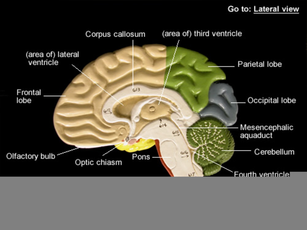
Human Brain: Structure, Location, Function, Parts & Pictures The human brain is the main Central Nervous System organ, situated in the head, protected by the cranium. Human brain has the same overall construction and anatomy as other mammalian brains, but it has a more developed cerebral cortex. The human brain is particularly complex and extensive. It embodies 2% of body mass, but it takes approximately ... Labeled Diagrams of the Human Brain You'll Want to Copy Now The central core consists of the thalamus, pons, cerebellum, reticular formation and medulla. These five regions are the central areas that regulate breathing, pulse, arousal, balance, sleep and early stages of processing sensory information. The thalamus interprets the sensory information and helps determine what is good and bad. Brain Label - The Biology Corner Image of the brain showing its major features for students to practice labeling. Answers are included. Brain - Human Brain Diagrams and Detailed Information Stria Medullaris of Thalamus Thalamus (3rd Ventricle) Tuber Cinereum Cerebrum Angular Gyrus Anterior Commissure of Brain Calcarine Sulcus Central Sulcus of Rolando Cingulate Gyrus Cingulate Sulcus Corpus Callosum Cuneus Fornix of Brain Frontal Pole Inferior Frontal Gyrus Inferior Parietal Lobule Inferior Temporal Gyrus Lamina Terminalis
Biology: The Human Brain
Label the Human Brain - 4th Grade Science Worksheet - SoD Your brain helps your body to run smoothly and controls everything you do, asleep or wide awake. This science worksheet for 4th grade helps you learn about its different parts. Label the Human Brain - 4th Grade Science Worksheet - SoD
Frontal Bone of the Human Skull | ClipArt ETC
3D Brain This interactive brain model is powered by the Wellcome Trust and developed by Matt Wimsatt and Jack Simpson; reviewed by John Morrison, Patrick Hof, and Edward Lein. Structure descriptions were written by Levi Gadye and Alexis Wnuk and Jane Roskams .

Gyri and Sulci on the Brain | ClipArt ETC
The Human Brain Atlas at Michigan State University The Human Brain Atlas Keith D. Sudheimer, Brian M. Winn, Garrett M. Kerndt, Jay M. Shoaps, Kristina K. Davis, Archibald J. Fobbs Jr., and John I. Johnson Radiology Department, Communications Technology Laboratory, and College of Human Medicine, Michigan State University; National Museum of Health and Medicine, Armed Forces Institute of Pathology
27 Label The Brain Anatomy Diagram - Wiring Database 2020
Brain Anatomy and How the Brain Works - Hopkins Medicine Gray and white matter are two different regions of the central nervous system. In the brain, gray matter refers to the darker, outer portion, while white matter describes the lighter, inner section underneath. In the spinal cord, this order is reversed: The white matter is on the outside, and the gray matter sits within.

Brain Diagram - Labeled - Tim's Printables
5 Human Brain Labels Stock Photos - Dreamstime Browse 5 professional human brain labels stock photos available royalty-free. Reset All Filters. Parts of the human brain and the functions for each part. In the background there is a monitor and keyboard. Concept for brain functions and activity. Parts of the human brain and the functions for each part. In the background there is a monitor and ...

TYWKIWDBI ("Tai-Wiki-Widbee"): Shark brain vs. dolphin brain
Brain (Human Anatomy): Picture, Function, Parts, Conditions, and More • The cortex is the outermost layer of brain cells. Thinking and voluntary movements begin in the cortex. • The brain stem is between the spinal cord and the rest of the brain. Basic functions like...

Diagram of Human Brain in Vertical Section | ClipArt ETC
40+ Diagram Of The Brain With Labels And Functions Pics Colored And Labeled Human Brain Diagram Stock Illustration … from media.istockphoto.com. All the functions are carried out without a single glitch and before you even bat an eyelid. Diagram of the eye muscles label brain worksheet labeling. To list all the functions and responsibilities of this collection of billions of neurons, you need to ...

Human Anatomy Lab: Heart Models
Diagram Of Brain with their Labelings and Detailed Explanation The parietal lobe is found at the upper back of our brain. This lobe functions by controlling all our complex behaviours, including senses of vision, the sense of touch, spatial orientation and body awareness. It manages body position, movements, the perception of stimuli, orientation, handwriting and visuospatial processing. The Occipital Lobe
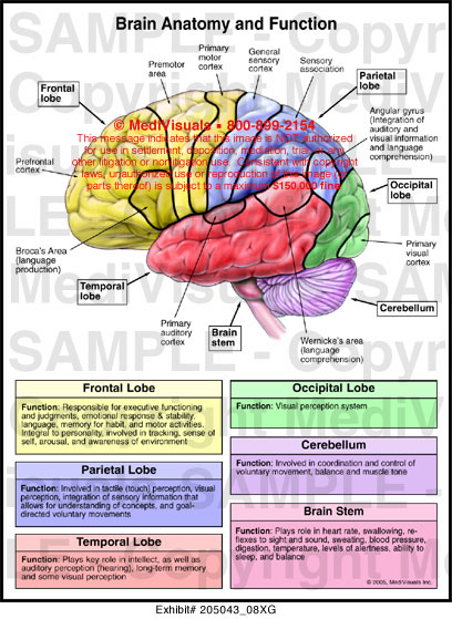
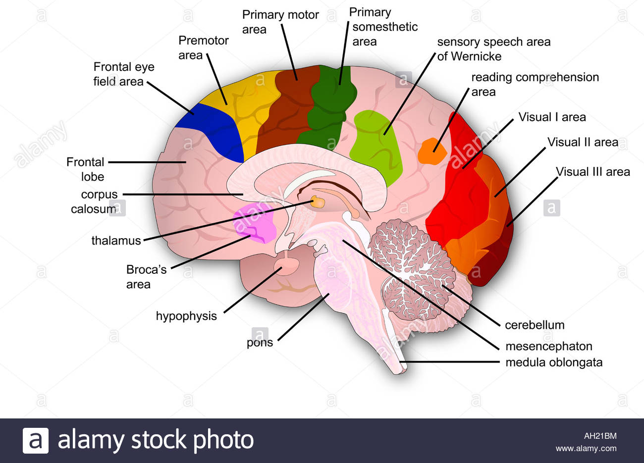
Brain Anatomy and Function Medical Illustration Medivisuals
Human Brain Anatomy - Components of Human Brain with Images Composed of the right and left hemispheres, it is the largest part of the brain and is responsible for the processing of speech, learning, reasoning, emotions, muscular contractions as well as the interpretation of sensory data related to hearing, vision and touch. ii. Cerebellum—the Sub-Cerebral Region:
Labeled Brain Model | Free Images at Clker.com - vector clip art online ...
Label The Brain - Mr. Barth's Class You won't label the parts of the brain on this website, but you'll familiarize yourself with the location of the parts and their basic functions. Lobes of the Brain Click on the link to the left to label the lobes of the brain. See how quickly you can do it with 100% accuracy. Lobes and Neuron Diagram

WMU Psychology Department: Lisa Baker
Human brain - Wikipedia The brainstem includes the midbrain, the pons, and the medulla oblongata. Behind the brainstem is the cerebellum ( Latin: little brain ). The cerebrum, brainstem, cerebellum, and spinal cord are covered by three membranes called meninges. The membranes are the tough dura mater; the middle arachnoid mater and the more delicate inner pia mater.

Diagram Unlabeled Brain Anatomy - Aflam-Neeeak
Frontiers | 101 Labeled Brain Images and a Consistent Human Cortical ... Labeling the macroscopic anatomy of the human brain is instrumental in educating biologists and clinicians, visualizing biomedical data, localizing brain data for identification and comparison, and perhaps most importantly, subdividing brain data for analysis.

Brain Viewed from Above | ClipArt ETC

human brain labeled - Google Search | Zoology | Pinterest | The o'jays ...
Anatomy Of The Brain Poster Chart Human Educational Poster #1 | A4 A3 ...

33 Human Brain With Label - Labels Database 2020

1000+ images about Brain graphics, photos & Art on Pinterest | Maze ...
Post a Comment for "42 labels of the human brain"